A heart ultrasound sees what a blood test and an EKG cannot: the heart itself, moving in real time. It shows how well the heart pumps, whether its valves are working, and whether its chambers have quietly changed shape under years of strain. Much of what it finds produces no symptoms until it becomes serious, which is exactly why it belongs in a conversation about workforce health. And because it uses sound waves rather than radiation, it travels well, making it one of the more practical advanced cardiac tests to bring directly to a workplace. Here is what a heart ultrasound reveals, and why employers are increasingly bringing it on-site.
Key Insights
- A heart ultrasound images the heart in motion: An echocardiogram uses sound waves to show the heart's structure and pumping action in real time, revealing things a blood panel or EKG cannot.
- It measures how well the heart pumps: The ejection fraction, the share of blood pushed out with each beat, is a key indicator of heart function, with a normal range generally around 50 to 70 percent.
- It catches silent structural problems: Valve disease, an enlarged or thickened heart, and reduced pumping strength often develop without symptoms and show up clearly on an echo.
- It uses no radiation: Because it relies on ultrasound, a heart ultrasound is safe, non-invasive, and repeatable, which also makes it practical to perform outside a hospital.
- On-site delivery removes the access barrier: Bringing the test to the workplace reaches employees who would never schedule an advanced cardiac test on their own.
What a Heart Ultrasound Actually Is
The name sounds more complicated than the test. A heart ultrasound is simply an ultrasound aimed at the heart.
An Ultrasound for the Heart
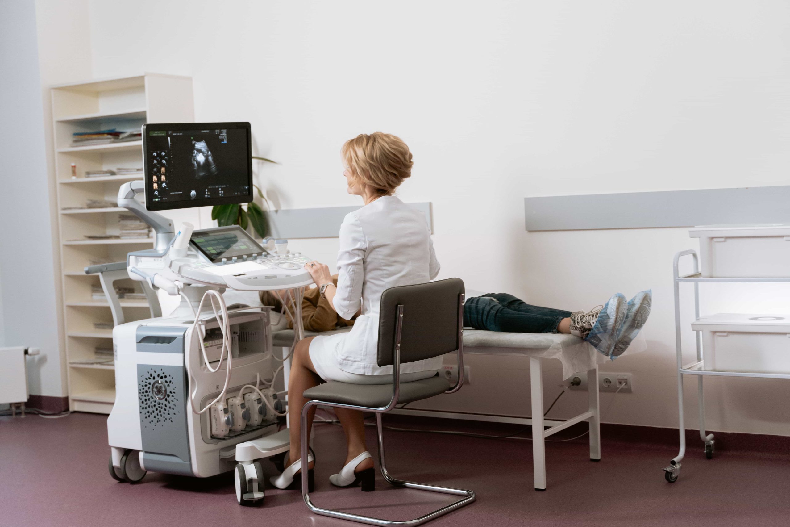
A heart ultrasound, known medically as an echocardiogram or echo, uses high-frequency sound waves to create moving images of the heart. The most common version, a transthoracic echocardiogram, is performed by moving a small probe across the chest with gel, the same basic technology used for prenatal imaging. It is painless, non-invasive, and involves no radiation, and it produces a real-time view of the heart beating.
How It Differs From an EKG
People often confuse the two, but they answer different questions. An EKG records the heart's electrical activity and rhythm. A heart ultrasound shows the heart's physical structure and how well it pumps. One reads the electrical signals, the other looks at the muscle, chambers, and valves in action. Because they reveal different things, they are often used together as part of a fuller cardiac picture.
What a Heart Ultrasound Reveals
This is where the test earns its value. A single scan produces a remarkable amount of information about how the heart is actually working.
Pumping Strength and Ejection Fraction
One of the most important measurements is the ejection fraction, the percentage of blood the left ventricle pushes out with each beat. A normal ejection fraction generally falls around 50 to 70 percent. A number below that range can indicate a weakened heart muscle and is a key marker used to diagnose and monitor heart failure and cardiomyopathy. It is one of the clearest single readings of how well a heart is doing its job.
Valve Function
The heart's four valves act as one-way doors that keep blood moving in the right direction. An echo can reveal stenosis, where a valve has narrowed, and regurgitation, where a valve leaks backward. Doppler imaging, included in many echo exams, measures the speed and direction of blood flow to gauge how serious a valve problem is. Valve disease often progresses silently, which makes finding it early genuinely valuable.
Chamber Size and Wall Thickness
The scan also shows the size of the heart's chambers and the thickness of its walls. An enlarged chamber or a thickened wall can be the physical signature of long-standing high blood pressure, heart failure, or a valve problem. These changes accumulate quietly over years, and an echo makes them visible.
Fluid, Structure, and More
An echo can also detect fluid around the heart, certain structural and congenital conditions, and how well the heart relaxes and fills between beats. Together these findings build a detailed picture of cardiac health that no blood test can provide on its own.
What a Heart Ultrasound Does Not Do
Being clear about a test's limits is part of using it well, and the heart ultrasound has them.
It Images the Heart, Not the Coronary Arteries Directly
A standard echo shows the heart's structure and function, but it does not directly image the coronary arteries the way an angiogram or CT scan does. It can reveal the consequences of a blood-supply problem, such as a section of muscle that moves abnormally, but it is not a direct look at arterial blockages. That is why an echo works best as one part of a broader cardiac assessment, alongside an EKG, blood work, and a physician's evaluation, rather than as a standalone answer.
Why This Test Travels Well
Of all the advanced cardiac tests, the heart ultrasound is among the best suited to leaving the hospital and coming to a workplace.
Non-Invasive and Radiation-Free
Because an echo uses sound waves rather than radiation or injected contrast, it carries no radiation exposure and requires no special facility. Modern ultrasound equipment is portable, which means a capable operator can perform a quality scan in a mobile clinic or a private room on-site, not only in a hospital imaging suite.
Quick and Comfortable
A standard heart ultrasound is painless and does not require recovery time, so an employee can return to work immediately afterward. That combination of safety, portability, and convenience is exactly what makes it realistic to deliver advanced cardiac imaging in a workplace setting.
Why Employers Bring It On-Site
The practical case for on-site heart ultrasound comes down to a simple truth: the test only helps the people who actually get it.
Reaching People Who Would Not Otherwise Go
Advanced cardiac imaging is not something most employees seek out on their own. It requires a referral, an appointment, and time away from work, and healthy-feeling people rarely pursue it. Bringing the test on-site removes every one of those barriers. The employees most at risk, and least likely to schedule a cardiac test themselves, get access during a normal workday.
Finding Silent Disease Early
Much of what a heart ultrasound detects, weakened pumping, valve disease, an enlarged heart, produces no symptoms until it is advanced. For a workforce, that means real cardiac problems can be identified while they are still manageable rather than after they become emergencies. Pairing an echo with the rest of a cardiac screen gives employees a genuine picture of their heart health. Our look at how often employees actually need a check-up covers where screening like this fits into a wellness program.
How NDS Wellness Brings Heart Ultrasound On-Site
Heart ultrasound is core to what NDS Wellness does. As a mobile cardiology and radiology provider, we bring echocardiography directly to your workplace through our corporate wellness program, as part of a full cardiac assessment that also includes a 12-lead EKG and comprehensive blood work. Employees get advanced cardiac imaging without a referral, a separate appointment, or a trip to a hospital.
Because we arrive in a mobile wellness clinic equipped with onboard diagnostics, testing happens across every shift, so the whole team gets access. A physician reviews the images and results in person, explains what they mean, and coordinates follow-up when a finding needs attention. It is hospital-grade cardiac insight delivered where your people already are.